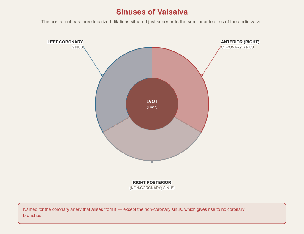
The aortic root consists of three localized dilations in the aortic wall situated just superior to the semilunar leaflets of the aortic valve: the left coronary sinus, the anterior (right) coronary sinus, and the right posterior (non-coronary) sinus.
Related RCIS terms
Study RCIS terms in context
Cardiac Codex turns definitions like this into a full study system: 179 lessons, 2,198 practice questions with explanations, four blueprint-matched full-length mock exams, live and timed, and Cardiac Coach — an AI tutor grounded in this exact glossary you can ask from any lesson.
Start free Back to the glossary →
Cardiac Codex is an independent study tool published by MdoubleA LLC, not affiliated with or endorsed by CCI or any certifying body. This glossary entry is exam-preparation information, not medical advice or a substitute for clinical judgment. Always verify current clinical practice against primary sources and institutional protocol.