Key takeaways
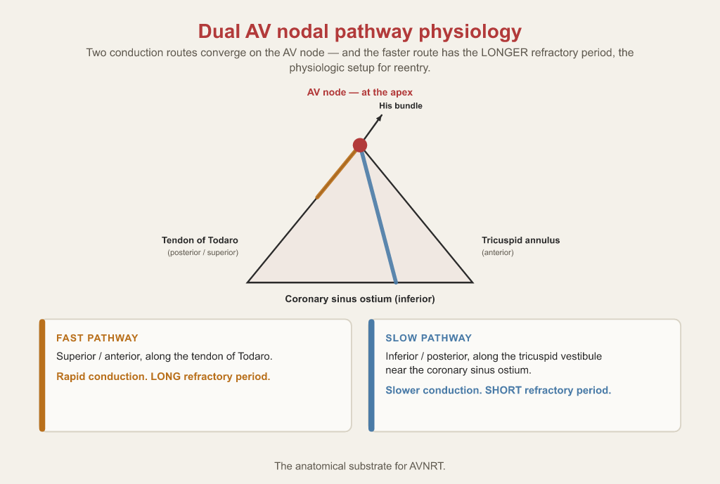
- The AV node sits at the apex of the triangle of Koch, bounded by the tendon of Todaro, the coronary sinus ostium and the tricuspid annulus.
- Dual AV nodal pathways (fast: rapid conduction, long refractory period; slow: slower conduction, shorter refractory period) are the substrate for AVNRT.
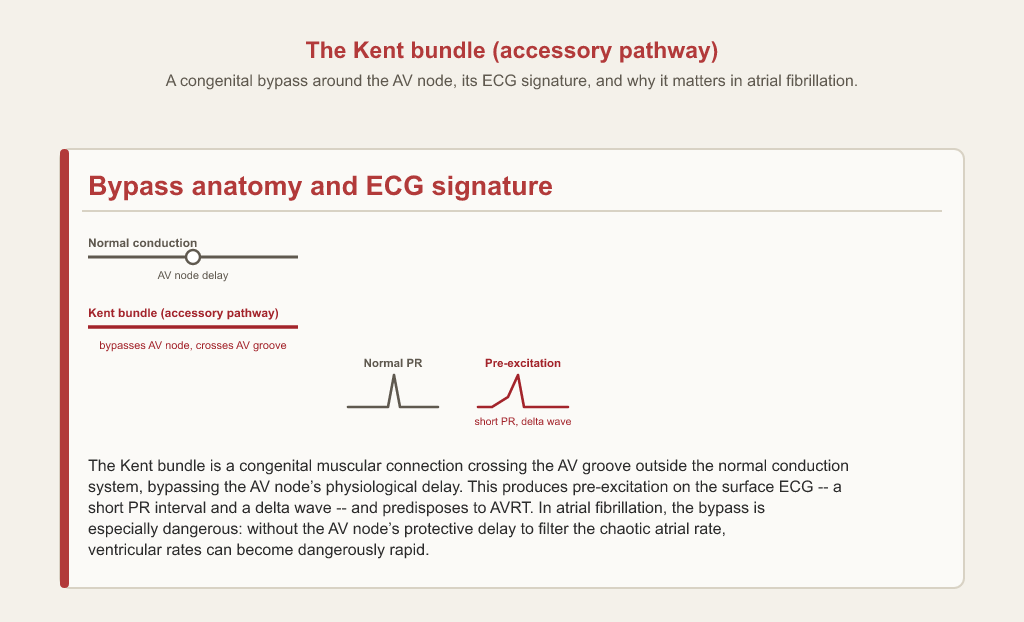
- A Kent bundle bypasses the AV node, producing a short PR and delta wave; in pre-excited atrial fibrillation, AV-nodal-blocking drugs can be dangerous.
- Mobitz I shows progressive PR lengthening before a dropped beat; Mobitz II drops a beat without prior PR prolongation and carries higher risk of complete block.
- Normal AH interval is 55–125 ms; normal corrected SNRT is generally under 525–550 ms in adults.
The short answer
The AV node sits at the apex of the triangle of Koch, and its dual fast and slow pathways are the substrate for AVNRT. An accessory pathway such as a Kent bundle bypasses it, causing pre-excitation. The EP study measures this system: the AH interval (normal 55–125 ms) and corrected sinus node recovery time are core baselines on the RCES.
The AV node and its two pathways
Most of what the EP study measures happens in or around the AV node, so start with where it sits and how it is wired.
Triangle of Koch
A right atrial region bounded by the tendon of Todaro, the coronary sinus ostium, and the tricuspid annulus, housing the AV node at its apex; the anatomical substrate for AVNRT given the AV node's dual fast and slow conduction pathways.
Dual AV Nodal Pathway Physiology
The fast pathway (superior/anterior along the tendon of Todaro, rapid conduction, long refractory period) and slow pathway (inferior/posterior near the CS ostium, slower conduction, shorter refractory period) within the AV node, forming the anatomical substrate for AVNRT.
Accessory pathways and pre-excitation
An accessory pathway is the other classic substrate: a connection that skips the AV node entirely.
Kent Bundle (Accessory Pathway)
A congenital muscular connection crossing the AV groove outside the normal conduction system, bypassing the AV node's physiological delay; causes pre-excitation (short PR, delta wave) and predisposes to AVRT and, in atrial fibrillation, dangerously rapid ventricular rates.
WPW / pre-excitation
A short PR interval combined with a slurred initial upstroke of the QRS (a delta wave) reflects early ventricular activation through an accessory pathway bypassing the normal AV nodal delay; recognizing this pattern matters procedurally because AV-nodal-blocking drugs (calcium channel blockers, in some cases adenosine) can be dangerous if the patient develops pre-excited atrial fibrillation, since blocking the AV node preferentially can promote even faster conduction down the accessory pathway.
When conduction fails: heart blocks
The surface ECG patterns of AV block are the bradycardia side of the same system.
Heart blocks
First-degree AV block shows a prolonged but constant PR interval with every P wave conducted; second-degree Mobitz I (Wenckebach) shows progressively lengthening PR intervals until a beat is dropped; Mobitz II shows a sudden dropped QRS without preceding PR prolongation and carries higher risk of progression to complete block; third-degree (complete) heart block shows complete dissociation between P waves and QRS complexes, each marching to its own independent rate.
The EP study: what gets measured
The electrophysiology study puts numbers on the conduction system's behaviour. Two baseline measurements come up repeatedly.
Electrophysiology Study (EPS)
A procedure using percutaneous multipolar intracardiac catheters to systematically assess electrical stimulation, conduction velocities, and arrhythmogenic substrates of the myocardium, providing definitive diagnosis of tachyarrhythmias and bradyarrhythmias.
AH Interval
The portion of the baseline intracardiac conduction interval (normal 55-125 ms) denoting AV nodal conduction time, measured from the atrial to His bundle electrogram and highly susceptible to autonomic tone and decremental delay.
Sinus Node Recovery Time (SNRT)
The simplest pacing protocol, the S1 train, delivers fixed-rate pulses to test the automaticity of the SA node. To measure the SNRT, the HRA is paced at a rate slightly higher than the intrinsic sinus rate for a sustained period of 30 to 40 seconds. Cycle lengths typically range from 700 ms down to 300 ms across multiple attempts. Upon the abrupt cessation of pacing, the clinician measures the exact time it takes for the SA node to recover from overdrive suppression and generate its first spontaneous impulse. A prolonged SNRT is a primary indicator of sinus node dysfunction. To streamline this process across patients with different baseline heart rates, the corrected SNRT (CSNRT = SNRT minus baseline sinus cycle length) is calculated instead of the raw value. Normal CSNRT is generally <525-550 ms in adults; values above this threshold are considered abnormal and support a diagnosis of sinus node dysfunction.
How this shows up on the RCES exam
Diagnostic procedures are 26% of the RCES blueprint per CCI's published content outline, and AV-node function is part of that domain. Expect to name the substrate behind a tachycardia, read a baseline interval against its normal range, and recognise a block pattern from its PR behaviour.
Test yourself on this
The free 25-question diagnostic scores you by blueprint domain, so you can see whether this topic is where your points are leaking.
Start the free diagnostic Back to the exam guide →Frequently asked
What is the substrate for AVNRT?
Dual AV nodal pathway physiology: a fast pathway with rapid conduction and a long refractory period, and a slow pathway with slower conduction and a shorter refractory period, within the triangle of Koch.
What is a normal AH interval?
55 to 125 ms. It reflects AV nodal conduction time, measured from the atrial to the His bundle electrogram, and is highly susceptible to autonomic tone.
How is corrected sinus node recovery time calculated?
CSNRT is the SNRT minus the baseline sinus cycle length. Normal is generally under 525 to 550 ms in adults.
Why are AV-nodal-blocking drugs dangerous in WPW?
In pre-excited atrial fibrillation, blocking the AV node can promote even faster conduction down the accessory pathway.
How do Mobitz I and Mobitz II differ?
Mobitz I (Wenckebach) shows progressively lengthening PR intervals until a beat drops; Mobitz II drops a QRS suddenly without preceding PR prolongation and carries a higher risk of progressing to complete block.