Key takeaways
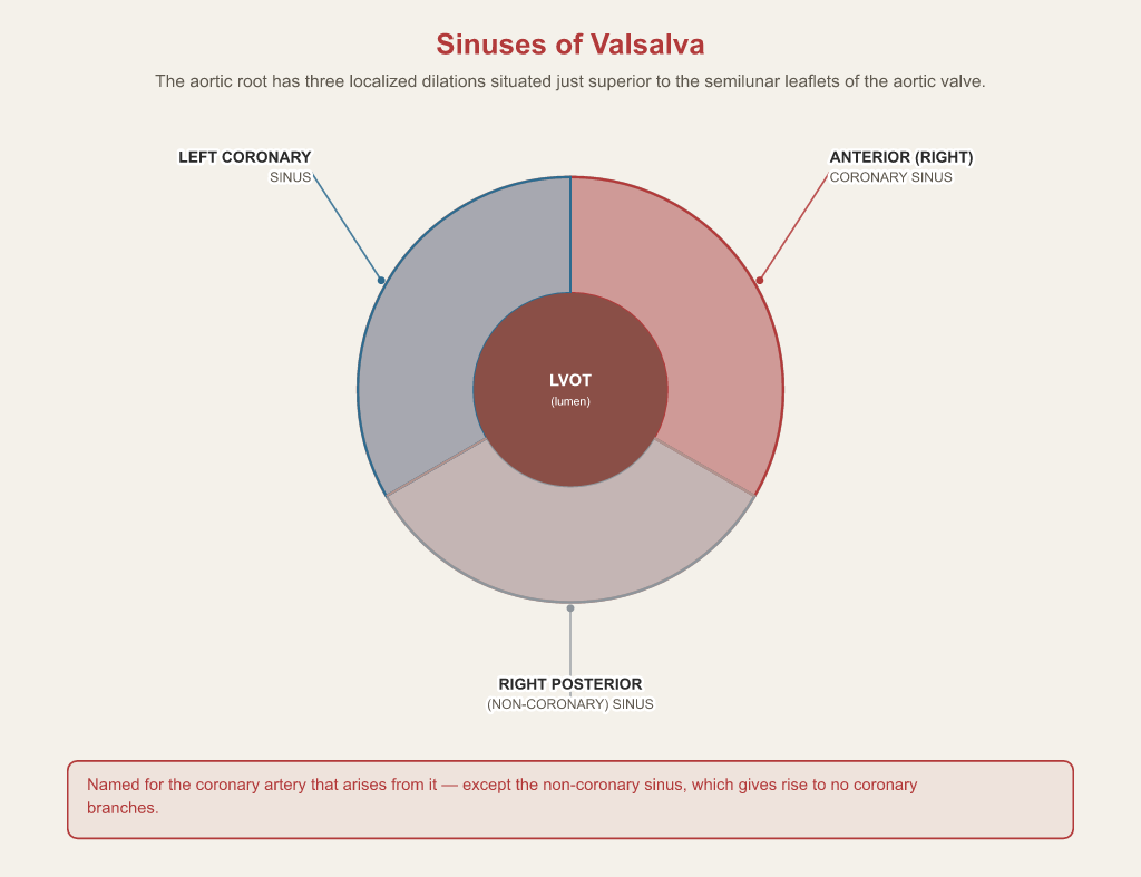
- The aortic root has three sinuses of Valsalva: left coronary, anterior (right) coronary, and right posterior (non-coronary).
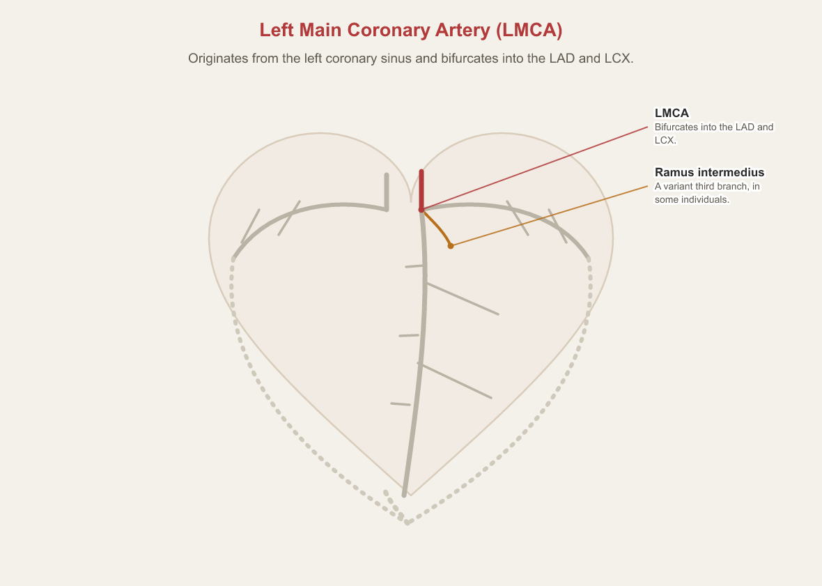
- The left main arises from the left coronary sinus and bifurcates into the LAD and the circumflex; some people have a third branch, the ramus intermedius.
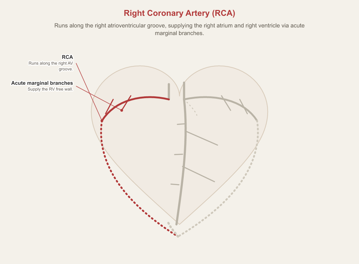
- The LAD gives off septal perforators and diagonals; the circumflex gives off obtuse marginals; the RCA gives off acute marginals.
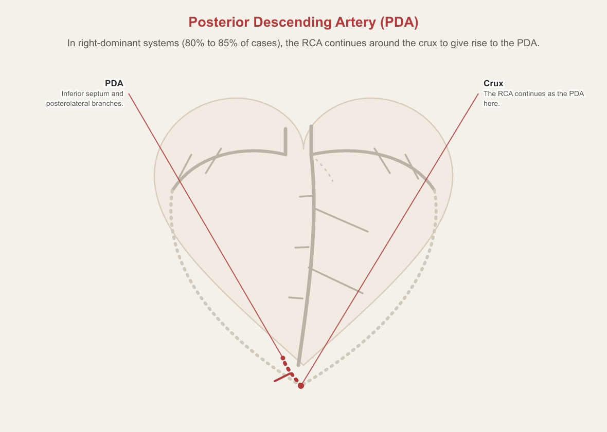
- In right-dominant systems (80–85% of cases) the RCA gives rise to the PDA.
- The RAO caudal ("spider") view is the optimal projection for the left main bifurcation, proximal LAD, circumflex origin and ramus.
The short answer
The left main arises from the left coronary sinus and divides into the LAD (septal and diagonal branches) and the circumflex (obtuse marginals). The RCA runs in the right AV groove and, in right-dominant hearts (80–85%), gives rise to the PDA. The RCIS exam also tests conduction-system blood supply and the projections that show each segment.
Where the coronaries start: the aortic root
Every coronary engagement starts at the aortic root, so the anatomy of the sinuses is the first thing to have straight.
Sinuses of Valsalva
The aortic root consists of three localized dilations in the aortic wall situated just superior to the semilunar leaflets of the aortic valve: the left coronary sinus, the anterior (right) coronary sinus, and the right posterior (non-coronary) sinus.
The left system
The left coronary system starts as a single short trunk and divides almost immediately.
Left Main Coronary Artery (LMCA)
Originates from the left coronary sinus and bifurcates into the Left Anterior Descending (LAD) and Left Circumflex (LCX) arteries. In some individuals, a third branch—the ramus intermedius—arises at this junction.
Left Anterior Descending (LAD) Artery
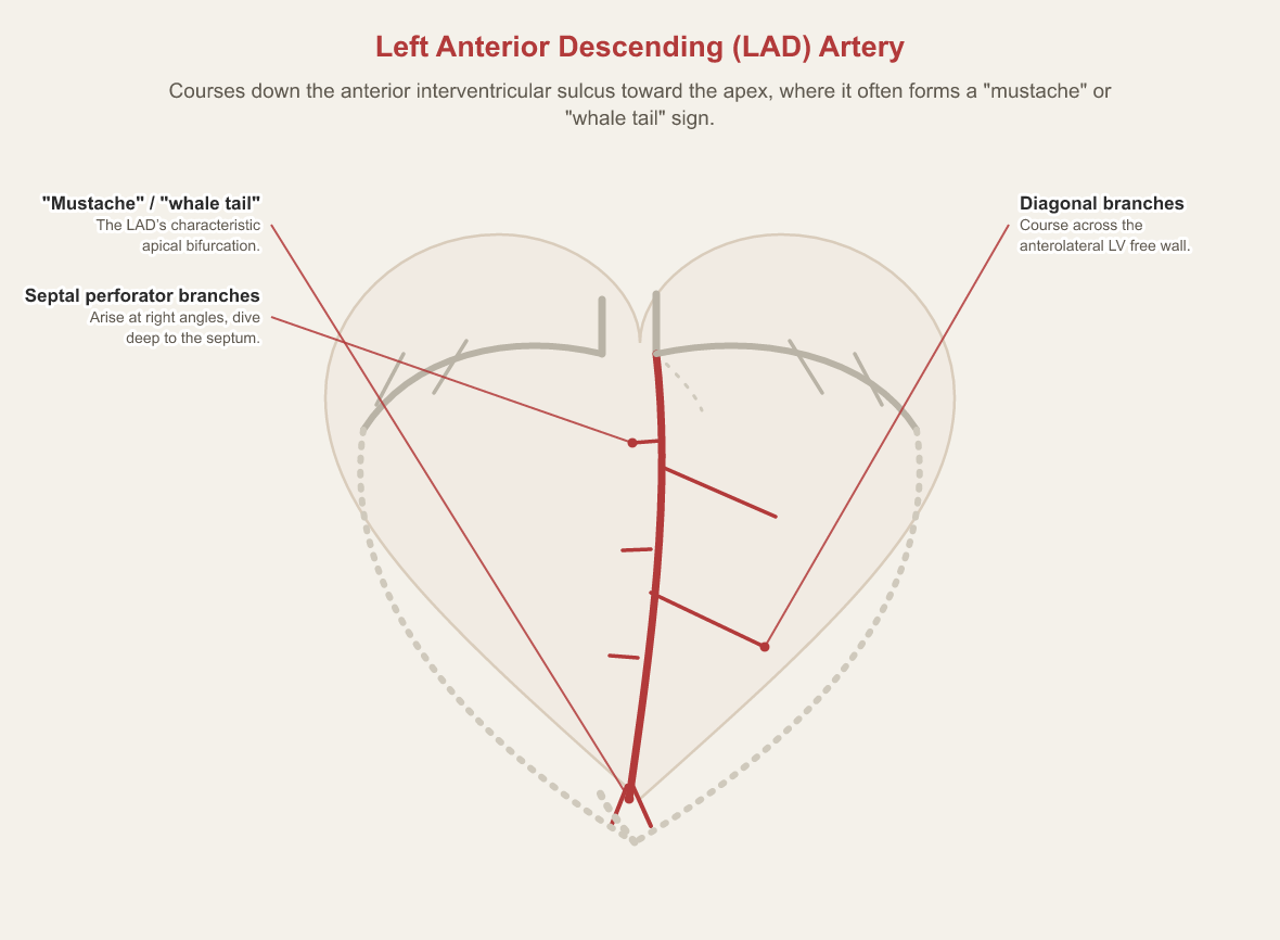
Courses down the anterior interventricular sulcus toward the cardiac apex, where it frequently branches in a characteristic "mustache" or "whale tail" sign. It gives off septal perforator branches, which arise at right angles and dive deep to supply the interventricular septum, and diagonal branches, which course diagonally across the anterolateral free wall of the left ventricle.
Left Circumflex (LCX) Artery
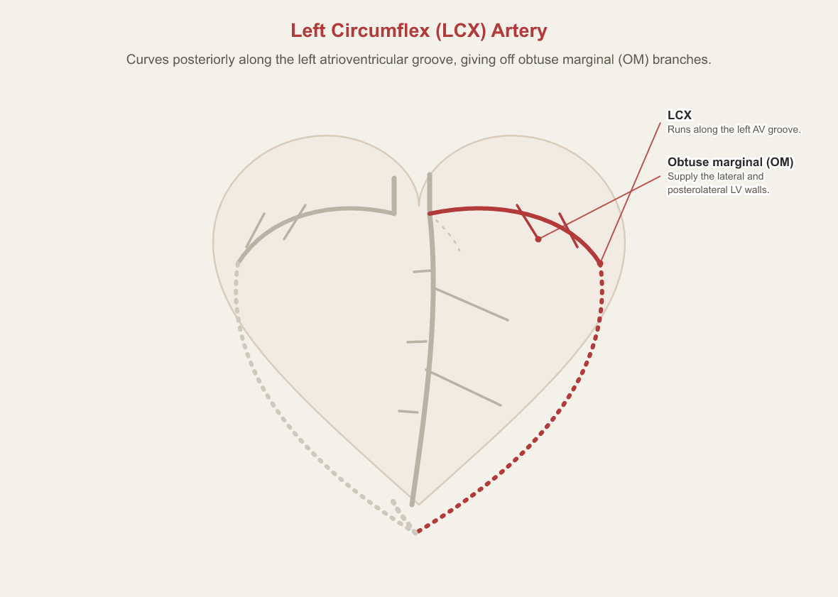
Curves posteriorly along the left atrioventricular groove, giving off obtuse marginal (OM) branches that supply the lateral and posterolateral walls of the left ventricle.
The right system and dominance
Dominance is defined by which artery supplies the posterior descending artery.
Right Coronary Artery (RCA)
Runs along the right atrioventricular groove, supplying the right atrium and right ventricle via acute marginal branches.
Posterior Descending Artery (PDA)
In right-dominant systems (80% to 85% of cases), the RCA continues around the crux of the heart to give rise to the PDA, which supplies the inferior third of the interventricular septum and posterolateral branches.
Blood supply to the conduction system
Knowing which artery feeds the conduction system explains why some occlusions produce rhythm disturbances. The figures below are the granular ones; as each entry notes, board-review sources often round them.
Sinoatrial (SA) Node
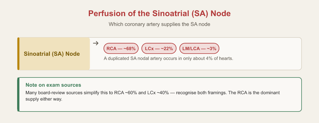
Supplied by the SA nodal artery, which arises from the RCA in roughly 68% of cases, the left circumflex in roughly 22%, and the left main/LCA proper in roughly 3%; a duplicated SA nodal artery occurs in only about 4% of hearts. Note: many board-review sources simplify this to RCA ~60% and LCx ~40%, so recognise both framings — the RCA is the dominant supply either way.
Atrioventricular (AV) Node
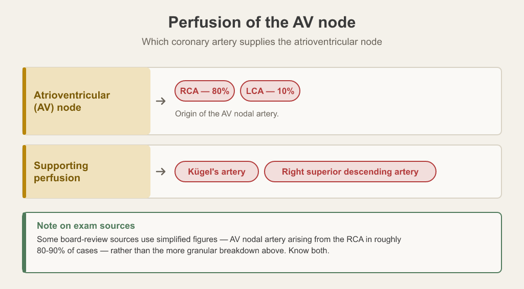
Perfused by the AV nodal artery, which typically arises from the RCA (80% of cases) or the LCA (10% of cases). Supporting perfusion is also supplied by Kügel's artery and the right superior descending artery. Note: some board-review sources use simplified figures -- AV nodal artery arising from the RCA in ~80-90% of cases -- rather than the more granular breakdown above.
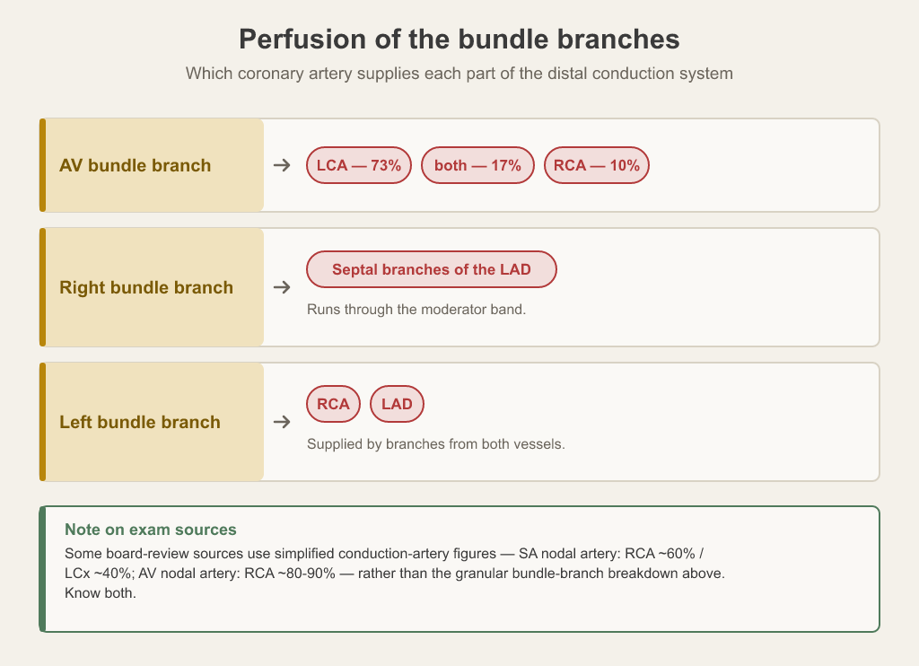
Bundle Branches
The AV bundle branch is supplied by the RCA (10% of cases), the LCA (73%), or both (17%). The right bundle branch (running through the moderator band) is supplied primarily by septal branches of the LAD. The left bundle branch is supplied by branches from both the RCA and LAD. *Note: some board-review sources use simplified conduction-artery figures (SA nodal artery: RCA ~60%/LCx ~40%; AV nodal artery: RCA ~80-90%) rather than the granular bundle-branch breakdown above.*
Seeing it on the screen
Anatomy on the exam is usually anatomy as it appears under fluoroscopy.
RAO Caudal ("Spider View")
The optimal fluoroscopic projection for visualizing the LMCA bifurcation, the proximal LAD, the origin of the LCX, and the ramus intermedius, achieved by tilting the detector toward the patient's right and feet.
How this shows up on the RCIS exam
Coronary anatomy underpins both of the RCIS exam's largest domains: diagnostic procedures (40%) and interventional procedures (34%), per CCI's published content outline. Expect questions that name a vessel from its course, identify which territory a lesion threatens, or choose the projection that best shows a segment.
Test yourself on this
The free 25-question diagnostic scores you by blueprint domain, so you can see whether this topic is where your points are leaking.
Start the free diagnostic Back to the exam guide →Frequently asked
What are the branches of the left main coronary artery?
The left anterior descending (LAD) and the left circumflex (LCX); in some people a third branch, the ramus intermedius, arises at the bifurcation.
What determines coronary dominance?
Which artery gives rise to the posterior descending artery. In right-dominant systems, 80% to 85% of cases, it is the RCA.
Which artery supplies the AV node?
The AV nodal artery, which typically arises from the RCA (80% of cases) or the LCA (10%), with supporting perfusion from Kügel's artery and the right superior descending artery.
What is the spider view?
RAO caudal: the optimal projection for the left main bifurcation, the proximal LAD, the origin of the circumflex, and the ramus intermedius.